Damage to brain blood vessels seen in nearly half of those with TTP type
Researchers say imaging scans may help determine long-term risk
Written by |

For one-quarter of immune-mediated TTP patients, damage to the brain's blood vessels progressed desite remission. (Image from iStock)

Nearly half of people with immune-mediated thrombotic thrombocytopenic purpura (TTP) showed signs of damage to small blood vessels in the brain — known as cerebral small vessel disease, or cSVD — during an acute episode, a new study from Italy found.
Such damage, which poses a risk for stroke and cognitive decline, is detectable on MRI scans, according to the researchers, who sought to assess the prevalence and one-year progression of cSVD among people with this type of the rare bleeding disorder, known as iTTP. The team noted that cSVD was found in more than 45% of individuals with the disease, but in fewer than 30% of study controls, or people without iTTP.
Further, brain imaging scans over time showed that damage to these small blood vessels progressed in more than one-quarter of the iTTP patients in the following year, despite their disease being in remission.
“These findings indicate that cSVD is common during acute iTTP and progresses during the first year despite … remission,” the researchers wrote.
The results also “support MRI-based assessment of cSVD as a potential tool for long-term risk stratification in iTTP,” the team noted.
The study, “Cerebral Small Vessel Disease in Immune-Mediated Thrombotic Thrombocytopenic Purpura Patients During the Acute Phase and Disease Remission,” was published in the American Journal of Hematology.
iTTP occurs when the immune system mistakenly attacks ADAMTS13, an enzyme that helps regulate blood clotting. Without sufficient ADAMTS13 activity, small blood clots can form throughout the body, blocking blood flow and damaging organs, including the brain.
Although treatment can bring the disease into remission, patients may continue to experience neurological problems, which typically affect cognition and increase fatigue.
Scientists use MRI to look at blood vessel damage in brain
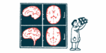
Considered common but not well-understood, cSVD affects small blood vessels in the brain. It can be detected on MRI brain scans as tiny areas of bleeding, damage to the brain’s white matter, or enlarged spaces surrounding blood vessels.
These changes are associated with a greater risk of stroke and cognitive decline. However, how frequently they occur in people with iTTP — and whether they worsen after an acute episode — has remained unclear.
Now, researchers at a hospital in Milan aimed to address this knowledge gap with a study involving 25 adults with iTTP. These patients had a median age of 56 and 60% were women. As controls, the team included 26 people without any known neurological disease.
The patients all underwent brain MRI scans during hospitalization, a median of seven days after their acute episodes began. Follow-up scans were performed in 22 patients approximately one year later, after they had achieved remission.
The researchers evaluated four MRI markers of small vessel damage and assigned each participant a score ranging from zero, for no detectable damage, to four, indicating all four types of damage.
7 times higher risk seen for iTTP patients in remission
During acute iTTP episodes, at least one sign of cSVD was detected in 48% of patients. That compared to one sign of such damage in 27% of controls.
Moderate small vessel damage was observed in 24% of the patients but in none of the controls. Tiny brain bleeds were the most common cSVD feature, affecting 32% of patients.
Additionally, 13 patients (52%) had MRI evidence of acute brain injuries caused by reduced blood flow. Most (92%) also showed neurological symptoms during their acute episode, the researchers noted.
A total of 22 patients underwent a follow-up MRI at a median of 12.5 months after the acute episode. The prevalence of cSVD increased from 50% during the acute episode to 64% during remission, the team noted.
The proportion of patients with moderate or more extensive damage also increased, from 27% to 41%, according to the researchers. Overall, six patients (27%) showed progression in their small vessel damage scores, even though all had achieved clinical and ADAMTS13 remission. No new acute injuries caused by reduced blood flow were detected on the follow-up scans.
After accounting for age and sex, the researchers determined that patients assessed during remission had a sevenfold higher risk of greater small vessel damage than the controls.
The scientists also explored whether lower ADAMTS13 activity during remission was associated with brain damage. Although patients whose damage progressed spent somewhat more time with lower enzyme activity, no clear association was established.
Study limitations noted by the investigators included the small number of participants and its single-center design. The team also noted that cardiovascular risk factors, such as hypertension (high blood pressure), were not matched in patients and controls.
“Future prospective studies should determine whether cSVD burden predicts long-term neurological and cognitive outcomes,” the researchers concluded.